|
01/16/2015 08:00 AM EDT
Blood agar plate culture of Corynebacterium haemolyticum.
|
| |
|
01/16/2015 08:00 AM EDT
Clostridium botulinum growing on egg yolk agar showing the lipase reaction after 72 hours of incubation. C. botulinum is a strictly anaerobic bacterium that when grown on egg yolk agar, its colonies will exhibit a lipase reaction, described as the shiny area around each colony.
|
| |
|
01/15/2015 08:00 AM EDT
This photomicrograph demonstrates cellular changes associated with rabies encephalitis using an H&E staining technique.
|
| |
|
01/14/15 08:00 AM EDT
This image shows Centers for Disease Control and Prevention (CDC) scientist Carol Bolden examining microscopic slides showing Exserohilum rostratum (see on computer screen) during the 2012 multistate fungal meningitis outbreak investigation.
|
| |
|
01/13/15 08:00AM EDT
Magnified 1000X, this photomicrograph depicts a number of Trypanosoma lewisi flagellate parasites that were present in a blood film specimen, and which had been highlighted using a Giemsa stain.
|
| |
|
01/12/15 08:00AM EDT
Prepared using the modified Steiner silver stain method, this photomicrograph revealed the presence of Treponema pallidum spirochetes in this tissue sample.
|
| |

 0001567
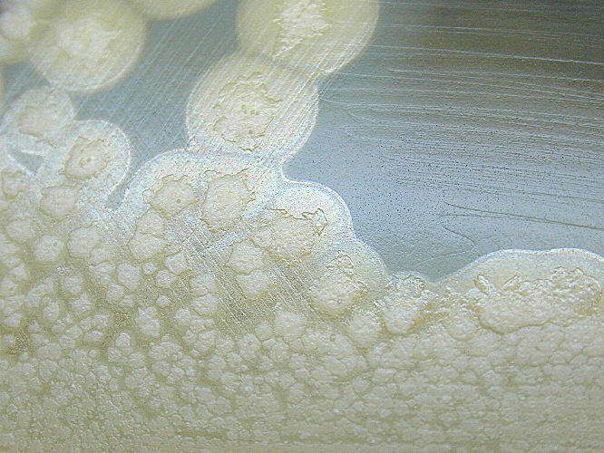 0001930
 0003981
 0015148
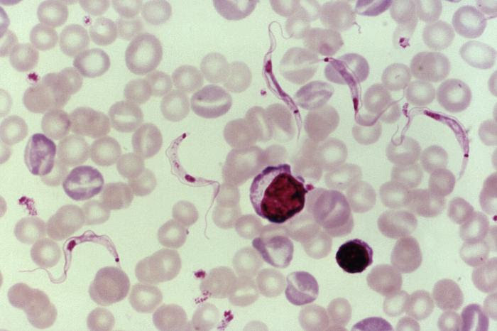 0014821
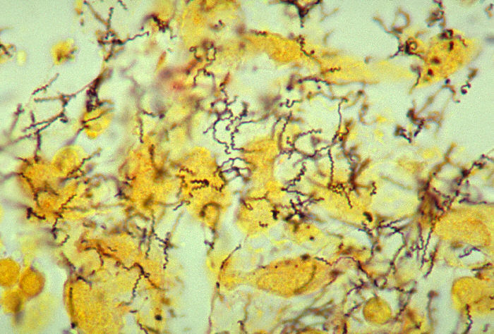 0000836
댓글 없음:
댓글 쓰기